|
Conversations with Neil’s Brain The Neural Nature of Thought & Language Copyright 1994 by William H. Calvin and George A. Ojemann. You may download this for personal reading but may not redistribute or archive without permission (exception: teachers should feel free to print out a chapter and photocopy it for students). William H. Calvin, Ph.D., is a neurophysiologist on the faculty of the Department of Psychiatry and Behavioral Sciences, University of Washington. George A. Ojemann, M.D., is a neurosurgeon and neurophysiologist on the faculty of the Department of Neurological Surgery, University of Washington. |
|
Losing Consciousness
NEIL LOSES CONSCIOUSNESS only occasionally during his seizures, and then only for a few
minutes. But back when he was first injured fifteen years ago, he was unconscious for a long
time. His head injury had damaged his brain stem. In a family letter written while he was still
recovering, his wife, Judy, described what happened:
But even aware has its problems. In the early days after his head injury, Neil was probably unaware of his surroundings — but we don’t really know that. He might have been aware of the voice and the pinch, but unable to respond; because he also had memory problems, maybe he couldn’t report that later. Occasionally, one encounters a patient who can recall conversations she overheard at a time when everyone thought she was still in coma. She was aware but effectively paralyzed. And so, rather than consciousness or awareness, neurologists prefer to talk about something they can objectively measure: levels of arousibility. A sleeping person can usually be aroused to full alertness, just by a loud voice. But sometimes the arousal achieves only a level of stupor, even when a pinch is used. And sometimes no purposeful movements result, in which case we talk of deep coma. Arousal is not the same as attention, another aspect of consciousness. Arousal is general, not specific like attention. The errors that people make during overarousal tend to be errors of commission or over-correction — they jump to conclusions. In under-arousal, one tends to get errors of omission instead — things aren’t noticed as they should be. Vigilance tasks, such as the sentry or the radar operator trying to stay alert for the rare event, lead to loss of arousal within times as short as a half hour. But that’s quite different from fatigue, which is commonly due to overarousal. Stress is, in some sense, overarousal. Levels of arousal may not be included in those consciousness connotations in Webster’s, but that’s what neurologists usually mean when they use the word. When physicians are forced to use the “C-word,” that’s the connotation that they will usually retreat to, the only firm ground in the whole morass. But equating conscious with arousable creates appalling problems. It tends to be interpreted as ascribing consciousness to any organism that has irritability. And irritability is a basic property of all living tissue, plant as well as animal, the first-trimester fetus as well as the severely demented Alzheimer’s patient — even the bacterium in your gut is conscious by that definition. A recent president of the United States used that definition in explaining his stance against abortion, although no reporter thought to ask him what he therefore thought of the consciousness of, say, a sperm. With so many major synonyms (aware, awake, deliberate, sensitive, arousable, and more), you can see why everyone gets a little confused talking about consciousness, the subtle thinkers as well as the literal-minded. Modern discussions of consciousness in the scientific community usually include such aspects of mental life as focusing your attention, things that you didn’t know you knew, mental rehearsal, imagery, thinking, decision making, awareness, altered states of consciousness, voluntary actions, subliminal priming, the development of the concept of self in children, and the narratives we tell ourselves when awake or dreaming.
 [FIGURE 8 The brain stem and arousal-attention mechanisms] |
|
NEIL’S STAGES OF RECOVERY correspond to more and more of his brain stem
recovering from the bruising it took during the sudden stop. The first good sign was when his
arms flexed, even though purposelessly; the second good sign was when he tried to push away
the hand that pinched him. Then he achieved stupor, and finally he was awake and observing his
surroundings. And now, fifteen years later, he is an experienced engineer asking lots of
questions about his brain. In the weeks before his surgery, I gave him much to read, and we met
in the hospital cafeteria to discuss it. Neil found me in the atrium one afternoon, in my favorite corner behind the espresso machine, just as a psychiatrist friend was leaving. So we had the round table to ourselves, amid the smell of fresh coffee and the sound of rain on the glass roof. It was where we’d met the previous week, when Neil had brought me the copy of the letter his wife had written fifteen years before, and it proved to be the table at which most of our pre-op conversations were held. After we had discussed his cab driver’s tales about other passengers she’d brought to this medical center, he asked about coma and consciousness. “So that’s just the `wake-up-call center’ down in my brain stem?” he asked. “Is coma merely deep sleep, where the brain’s alarm clock doesn’t work in the morning?” “Yes and no,” I answered. (Actually, I wasn’t that succinct, but I’m going to condense my answers a little). We know, from animal studies back in the 1940s, that there are certain parts of the brain stem that control awakening. They’re crucial. Animals with damage there appear to be continuously asleep and don’t awaken spontaneously. Many neurons — that’s what we call nerve cells — down there deliver norepinephrine all over the brain and spinal cord. And so diffusely that we think of it almost as a lawn-sprinkler system. A lot gets delivered to the sensory strip. About half of these “norepi” neurons are in a brain stem cluster known as the locus coeruleus. They are rather inactive during sleep, and most active when you startle at something unexpected. “I figured out that adrenaline is another name for epinephrine,” Neil commented. “I suppose that noradrenaline is just a synonym for norepinephrine?” Right, epinephrine does things outside the brain somewhat similarly to what norepi does inside. Adrenaline is released from the adrenal glands, which sit atop the kidneys, and is carried in the bloodstream as a hormone. When you get a rush from almost getting hit by a car, that’s adrenaline, raising your heart rate and setting up your muscles for fight or flight. It adds to the startle effect that norepi produces within the brain. There is another diffusely broadcasting group of neurons scattered along the centerline near the surface of the brain stem. These deliver serotonin to wide areas of the brain and spinal cord. The activity of these serotonin neurons may determine levels of arousal, but they don’t increase their activity with sensory input. The norepi neurons do, and are thought to be more important for the emphasis of attention. Now it looks as though the serotonin neurons have another big role, that of regulating pain perception. Drugs that enhance the effectiveness of norepinephrine and serotonin are commonly used as antidepressants. Ones that enhance the serotonin effects are often helpful in chronic pain disorders. During coma, neither the serotonin nor norepi systems are working well, which is probably why the patient can’t be aroused.
 [FIGURE 9 Stages of sleep, dreaming] |
|
BUT SLEEP IS FAR MORE COMPLICATED than coma, I continued. About two hours every
night is spent in light sleep. People awakened from that stage of sleep report mulling things over
but never getting very far along a line of thought. They’ll call this thinking, not dreaming.
In the deeper parts of this “orthodox” sleep is when bed-wetting occurs, as well as
night-walking episodes. Both norepi and serotonin neurons are ticking along at only about half
of their waking rates. About every 100 minutes or so during sleep, both the norepi and serotonin systems virtually shut down. Then the sleeper experiences an episode of “paradoxical” sleep. This is when most of the muscles are unusually relaxed — except (and here’s the paradox) for the eyes, which dart back and forth under the closed eyelids, and except (in males) for the penis, which becomes erect. If you wake up people in this stage of sleep, they’ll often report dreaming, not thinking. There are well-formed sensory impressions, though sometimes quite bizarre. A series of actions seems to occur, often with an improbable juxtaposition of people, places, and times. Strong emotions may be experienced. And the dreamer seems to accept, uncritically, what’s happening in the dream — memory recall doesn’t work very well in sleep. Making new memories is even more impaired. If you wait as much as five minutes after the end of the rapid eye movement sleep to awaken sleepers, they won’t remember much of the dream. Dreaming is mostly visual, with some auditory and tactile sensations as well as a sense of movement — but seldom does anyone report pain, or smells or tastes. “So they’re not really like daydreams, are they?” Neil asked, munching on a scone he’d bought at the espresso stand. People indulge in fantasies while awake, I told him, but these are seldom as vivid and bizarre as in nighttime dreams. Furthermore, real dreams are like delusions; you have very little insight, and while the dreams are occurring, you tend to think it’s all real. Emotions aren’t normal in dreams. Anxiety, fear, and surprise are likely to be exaggerated. Yet shame and guilt play little role in dreams, even when seemingly engaging in shabby behavior that would stir your conscience if you were awake. You don’t gain access to established memories very well during sleep, and you usually can’t establish new memories either. Actually, it’s a bit of a puzzle why you ever form permanent memories of dreams, given how poor the recall usually is. But there’s a way around that, at least if you awaken before the five minutes are up. If you recall a dream before it fades, when your circuits for memorizing new things are working once again, then you can memorize the recall rather than the original happening. If you make a habit of reviewing your fading dreams upon awakening, you can memorize quite a collection of nonsense. Keeping a dream diary can thus clutter up your head with a lot of things that didn’t really happen — and we have enough trouble as it is, just keeping straight those things that really happened from all those things that we merely imagined or planned. “So why do therapists like to talk about dreams so much?” Neil asked. Psychotherapists may find it useful to ask about dreams as a way of getting patients to talk about something other than their current problems, if they are dwelling on them. Talking about dreams can be a psychotherapist’s version of talking about the weather, simply a way of getting a conversation started so that it can move on to something that the patient would not think to mention spontaneously. A lot can be learned simply from the way a patient chooses to interpret a dream. But seeking significance in dreams — which is what patients may mistakenly think the therapist’s questions are all about — is not much better than seeking it in tea leaves. Randomness predominates in both, yet we usually try to make it fit into some sort of story line. “So, that’s the kind of sleep you can’t do without,” Neil went on. “You go a little crazy if you don’t get enough dreaming sleep, don’t you?” You normally dream about two hours every night, I told him, in addition to your two hours of mulling things over in light sleep. If you were awakened each time you started to go into dreaming sleep, you’d play catch-up the next night, dreaming more than normal. If this happened night after night, you’d become rather irritable — and not just because of awakening. You can be awakened just as often in orthodox sleep without becoming so cranky. “I read somewhere that, in dreams, we all have the experience of being psychotic or demented or delusional. It almost sounds like we have a quota for being psychotic, and if we don’t fulfill it at night, we become psychotic during the day!” That’s like what René Descartes said back in 1641: “I am accustomed to sleep and in my dreams to imagine the same things that lunatics imagine when awake.” Psychiatrists consider psychosis a little more complicated than that — but yes, dreams do show you normal physiological processes that, when nothing better is happening, could correspond to the symptoms of mental illness. There are people called narcoleptics, I went on, who suddenly fall asleep during the day. The problem was identified centuries ago, but we now know that narcoleptics go immediately into dreaming sleep — real daydreams, complete with the psychotic features of nighttime dreams. Usually dreaming sleep doesn’t occur until after at least twenty minutes of orthodox sleep, but in narcolepsy the transition can be very rapid. Indeed, the onset of the muscle relaxation accompanying dreaming sleep is so sudden in most narcoleptics that they may collapse while standing up. “I saw that happen to my roommate in the rehab hospital. Another survivor of seat belt neglect. I told him that he was taking `falling asleep’ too literally.” About three-quarters of narcoleptics sometimes do that, I said. Others get enough warning to lie down, or at least to rest their heads somewhere. Narcolepsy can be from a bruise to the brain stem, but most cases are probably inherited; there’s a “gene for narcolepsy” in 98 percent of narcoleptics. |
|
THE VARSITY CREW ROWED past us as we walked along the waterway behind the medical
center during a brief, sunny period. That’s when Neil brought up one of his vocabulary
questions. “I suspect you use the word stroke differently from the way the coxswain does,” he said. In medicine, I told him, the meaning of stroke was originally closer to “Felled by an unseen blow.” It meant any sudden collapse and loss of awareness. At least, that was the label for those cases in which the loss of consciousness wasn’t temporary, as in fainting or a seizure. It could, of course, have been a heart attack, but we now have a separate label for those cases of sudden collapse, since we understand them so well. So these days, stroke refers to what’s left over — sudden damage to the brain caused by blood-supply problems. Sometimes paralysis occurs, but it all depends on what brain region was damaged. Often no loss of consciousness occurs and the stroke manifestations are subtle. “What causes the stroke? The usual plumbing problems?” Leaks and stoppages, yes. And the occasional burst pipe can cause major structural damage rather quickly. Just as in a bruise under the skin, a blood vessel in the brain can leak. Blood is very toxic to neurons, which stop working and often die when it comes in direct contact with them. When there’s not an obvious external event like a hit on the head, bleeds are usually caused by the ballooning of an arterial wall, rather like an old tire that develops a blister on its sidewall. The balloon — called an aneurysm — may never cause any trouble — but occasionally it ruptures, pumping blood into the surrounding tissues. If a blood vessel in your leg were to rupture, the leg would just swell up. But the brain is surrounded by the skull, and all that escaped blood takes up space, squeezing the brain. That compression of the brain is what kills a lot of stroke victims. Or the stroke may be caused by a plugged-up blood vessel. Usually a clot forms, or an atherosclerotic plaque accumulating on an arterial wall comes loose — whichever, this embolus is swept along in the blood stream until it reaches an artery that is too small for it to pass through. Sometimes that unfortunate artery is in the lung, sometimes in the brain. It’s all pretty random, what artery becomes plugged. And so some region of the brain might lose its oxygen supply. If the obstruction is flushed through or dissolves with a short time, the neurological symptoms may be brief; one side of the face might sag, for example, but then begin to recover several minutes later. Such temporary strokes are known as transient ischemic attacks. But if the artery stays plugged up for something like fifteen minutes or more, permanent damage occurs. Once the dead cells are cleaned out, there’s a hole in the brain.
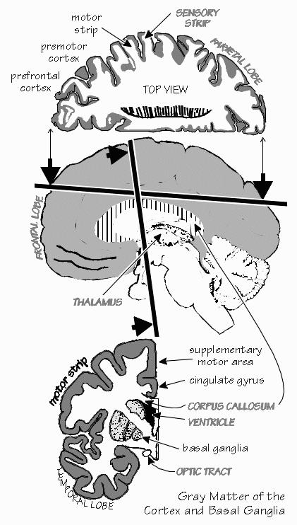
 [FIGURE 10 Gray matter of cortex and basal ganglia] “But I don’t have any holes in my brain, right?” Neil sounded a little anxious. Just the usual cavities that everyone has — those big reservoirs of cerebrospinal fluid we call the ventricles. There was a lot of excitement about the ventricles during the Renaissance. The first phrenologists were actually religious scholars who lived 500 years ago. They thought that the subdivisions of the soul were housed in the various cavities of the brain: memory in one; fantasy, common sense and imagination in another; thought and judgment in a third. “I’ll have a hole after the operation. What’s going to fill in the hole?” Cerebrospinal fluid fills all the space inside the skull that isn’t brain or blood. You’ll get an enlarged ventricle, so to speak. “I’ll have more soul, eh?” Neil chuckled as we walked back inside the cafeteria. “What about those people — I’ve seen them mentioned in magazine articles — who are discovered to have big holes in the heads — even though they have normal intelligence? They only seem to have a thin layer of brain.” Actually, most of their brain is normal, but they have a big, fluid-filled cyst, with only a thin layer of cerebral cortex in many places. People confuse the cerebral cortex with the brain in general, perhaps because the cortex is what seems to house the fancier functions such as language, planning ahead, and worrying about tomorrow. Back when the first computerized tomography scanners were being tested on medical students, one was discovered to have a big cyst like that, with only a thin layer of cortex. But that cortex worked pretty well. Of course, a thin layer of cortex is all that any of us has. It’s just a thin shell, weaving in and out of the folds in the brain’s surface. “In the pictures I’ve seen, it seems to vary in thickness from one place to another.” To explain that, I told him, would require that we get a piece of cake. With dessert in hand, I explained that most of that apparent variation in cortical thickness is just the cortex being sliced on an angle, rather than along the local vertical, just as if you took a layer cake and cut it on a slant. You can make it look two or three times thicker that way, by cutting on a really oblique angle. But in reality, the cortex is never any thicker than a stack of maybe two coins. It’s the icing on the cake. “Fooled me. So that thin little surface layer does all the interesting stuff? All the language? All the planning?” He thought for a minute. “So what’s the rest of the space between my ears, if there’s only a couple of thin layers of cortex doing all the work?” Wires. Lots and lots of wires. Except we call them axons or sometimes nerve fibers. If you’ve ever toured a telephone company’s central office, you’ll remember seeing huge bundles of wires rising out of the floor, continuing through the ceiling, and often ascending through several more floors of the building. But when you finally see the computer that switches all the calls from one circuit to another, you discover that it isn’t very large. It’s small enough to fit inside a closet. The brain does things much fancier than mere switching, but the wires still take up most of the room. That’s what the white matter is. The part of cerebral cortex doing all the fancy stuff is just that thin surface sheet, wrinkled into hills and valleys.
 [FIGURE 11 Cerebral cortex layers and wiretapping the pyramidal neuron] |
|
THE CEREBRAL CORTEX is an important proportion of the gray matter. But there is a lot
more in the depths of the brain, concerned with things like sleep, directing your attention, and
regulating your memory. Some gray matter, such as the thalamus, has an intimate back-and-forth
relationship with the cerebral cortex. Of course, the gray matter isn’t really gray. It’s closer to brownish red. “Gray matter isn’t gray anymore?” Neil said, laying down his fork in mock disgust. “My disillusionment is complete.” It’s only gray in a very dead brain. But then, that’s the only condition under which most people ever see a brain, assuming they ever see one at all. It’s only in the operating room that we get to see that the natural, workaday color of the gray matter is really a nice rich reddish brown — rather like color pictures of the Grand Canyon. “I hesitate to ask, but is the white matter really white?” Fear not, the white matter is indeed a pale porcelain white, not unlike skim milk diluted with a little water. The color is contributed by the fatty insulation, known as myelin, wrapped about the wires. There is some myelin insulation within the gray matter as well, but other things contribute some color too — all those other parts of the neuron, such as the cell body and dendrites, and lots of little blood vessels. In the white matter, you find only the long, thin, tubular part of the neuron — its axon — sometimes with myelin insulation wrapped around it, sometimes not. “Those wires carry the electrical signals, right? That’s where brain waves come from?” Yes and no. Yes, the axons carry electrical signals, called impulses, which are about a tenth of a volt in size and absolutely essential for moving information over long distances in cerebral cortex. But no — brain waves don’t come from axons in the white matter, but from another part of the cell — the dendrite — that is closer to the cortical surface. The electroencephalogram — EEG for short — is a pretty crude measurement. There’s nothing in the computer world that is as crude as the EEG. Still, it allows us to see little seizures happening that we otherwise wouldn’t know about, because they didn’t cause any muscles to contract. “But isn’t the EEG voltage proportional to the message traffic in some way — to the mail trucks arriving and leaving?” It’s not even as precise as those little tubes the traffic engineers lay across the street to measure traffic flow. But you don’t have to count every car and truck to get an idea of the traffic patterns. Just imagine using a tape recorder and hanging the microphone on a light pole. Set the tape speed very slow and let it run for a day, then watch the sound level indicator as you replay the tape. Do you know how you can wake up in a hotel room in a foreign city and guess whether it’s morning or not, without ever opening your eyes? If the traffic noise outside is just the occasional truck, it’s probably still four in the morning. Traffic starts to build about six, and so does the traffic noise. If you go back to sleep at six, you’ll probably wake up next during the height of the rush-hour noises.  [FIGURE 12 Neil’s EEG abnormality between seizures] The EEG shows some rhythms that led to the term “brain waves.” They are very much like the traffic rhythms you would notice. There are big, sustained peaks twice a day — during the morning and evening rush hours. There are more rapid rhythms too. If you plotted out the loudness of the traffic noise, you’d get a rhythm of about thirty little peaks every hour. “The traffic signals down the block probably cause the traffic to bunch up.” Right, I said. The rush-hour rhythm isn’t as pronounced on the weekends, and so there are cycles within cycles. The stop-and-go cycle becomes more pronounced during rush hours. So, too, the various EEG rhythms wax and wane in amplitude, depending on whether you are resting or alert, busy or waiting. The rhythms are very useful, even though quite imprecise. Sometimes we try to synchronize a lot of activity, just to see how the EEG responds. “That’s the test I had, with the clicks once a second, the one called the average evoked response?” Exactly. Just think of a loud noise — say, a dish breaking on the floor — that stops all the conversation in a room. Everything becomes unusually quiet. Then everyone begins talking at once. We can learn a lot about how some parts of the brain are working by using little disruptions like that. We can’t always figure out an appropriate stimulus to stop and restart the conversations. But for the visual cortex, light flashes work pretty well. |
|
NEIL WAS CURIOUS about his EEG abnormalities. He’s been though dozens of EEGs
over the years but never understood exactly what neurologists were looking for. We were
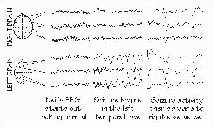
sketching some EEG patterns on the napkin he’d fetched from the espresso stand. “So what’s your analogy for the spike they find in my EEG? They said it’s the hallmark of a resting epileptic process, in between seizures.” The spike is more like a backfire — not normal, but not serious by itself. Usually an occasional backfire is just the sign of an engine that needs a tuneup, and I suspect that’s what most EEG spikes signify, too. It’s when you get a whole series of backfires, or a whole series of spikes in the EEG, that there’s a chance of a breakdown any minute. Sometimes you can see a series of spikes in the EEG that remind you of an old truck, surging ahead but bucking with another backfire, then surging again, threatening to shake itself to pieces with the repeated backfires, and finally all but collapsing in the street. That’s a lot like how we imagine a seizure starts.  [FIGURE 13 Neil’s EEG, as seizure begins] “How many neurons are involved in one of those EEG spikes?” Neil asked me. Probably millions, I replied. An EEG electrode glued to the scalp records signals from a region of brain at least the size of a dime. I once calculated that there are 133 million neurons in a dime-sized sheet of cerebral cortex. The troublemakers are probably only a small minority of that but, much of the time, their signal gets diluted by all the rest. A lot of seizures don’t produce muscular movements, yet they keep that part of the brain from getting any useful work done for a while. From the scalp, we can see only the larger mobs in action. When they reach the level of overt movements, they’re involving many millions of neurons. “Lots and lots of busy little neurons,” Neil said. “How many all told?” The count is now up to about 200 billion, I told him. That’s for the whole brain. For just the cerebral cortex, the subtotal is about 30 billion. People confuse the cerebral cortex with the entire brain, but the cortex has only a fraction of the total number of neurons. The cerebellum, atop the brain stem, has many more, thanks to so many little granule cell neurons. And, of course, the number of synapses is even larger — they’re the points of contact between neurons, and each neuron has many thousands of them. But even if the totals were constant across individuals, the subtotals would still vary between different parts of the cerebral cortex. If you were to look at the primary visual cortex in a number of normal individuals, you’d notice that it wasn’t always the same size. Some individuals have three times more than others. “So do those people see better?” |
|
IS BIGGER REALLY BETTER? In the case of the visual cortex, no one knows yet. Language
cortex probably varies a lot, too. Among various animal species, the depth of the cortical layers
varies somewhat, but the number of neurons beneath a square millimeter of cortical surface stays
quite constant, not far from 148,000. “That’s interesting. Sounds like some sort of packing principle is involved, that’s universal.” Perhaps. The major growth of cerebral cortex, as our ancestors became fancier and fancier primates, was sideways. In chimpanzees and gorillas, the total is less than a standard sheet of typing paper. Humans have the equivalent of four sheets of paper. Rats have less than a square inch of cortex, less than humans by a factor of 500. Increased surface area is presumably what led to so many wrinkles in the cortical surface, all those narrow valleys that increase the amount of surface area that can be packed into a given volume. “Does a smart person have more cerebral cortex than an idiot? Is cortical size what makes us smarter than a chimpanzee?” Cortical area has something to do with being smart, I conceded, although it’s hardly the whole story. If you measure the amount of gray matter using an MRI scan, individuals with high IQs have significantly more than those of average IQ. But the cortical area accounts for only a fraction of the variability in IQ. Something else must affect intelligence. The internal efficiency of the circuitry probably matters more than the total amount of cortex. “I’m going to lose some cortex when they do the surgery. What’ll happen to my IQ? My wife is concerned about that too. And I know my business partners are!” IQ measurements usually go up after operations like these, and rather significantly. But that’s because the patients are so impaired before the operation, either by the lingering effects of recent seizures or by high drug dosages and their side effects. And so individuals test lower than their biological endowment. Neil thought about this for a minute as we carried our trays over to the cafeteria’s conveyor belt. “So I’m likely to come out of this operation somewhat smarter than I am now — but not as smart as I was fifteen years ago before I banged up my head. But I was so smart back then that I didn’t wear my seat belt!” Just goes to show that good habits can be more important than IQ. Intelligence, or at least what IQ tests measure, has a lot to do with “being quick” mentally — able to get through a lot of questions in a fixed amount of time. And IQ also has a lot to do with being able to juggle many things at the same time, as in those analogy questions that ask “A is to B as C is to __?” And then you get three choices — D, E, and F — so that, to solve the problem, you have to keep six items in mind simultaneously. Things like judgment and creativity and a good storehouse of knowledge may be far more important in real life than whatever IQ measures. You don’t want to be so quick that you jump to the wrong conclusions, or so indecisive that you never make up your mind. It’s a matter of the right balancing act, what controls when you move on to the next problem. Having a “good brain” isn’t often a matter of how much brain there is. Quality can usually make up for quantity.
INSTRUCTORS: You may create hypertext links to glossary items in THE CEREBRAL CODE if teaching from Chapters 6-8, e.g., <a href=http://www.williamcalvin.com/bk9gloss.html#postsynaptic>Postsynaptic</a> |
 Conversations with Neil's Brain:
 The Neural Nature of Thought and Language (Addison-Wesley, 1994), co-authored with my neurosurgeon colleague, George Ojemann. It's a tour of the human cerebral cortex, conducted from the operating room, and has been on the New Scientist bestseller list of science books. It is suitable for biology and cognitive neuroscience supplementary reading lists. ISBN 0-201-48337-8. | AVAILABILITY widespread (softcover, US$12).
|